Atherosclerosis: From LDL Retention to Plaque and Thrombosis
Atherosclerosis is not simply "fat clogging a pipe."
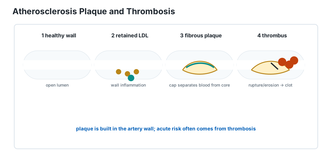
It is a chronic inflammatory disease of the artery wall involving retained cholesterol-containing lipoproteins, immune cells, smooth-muscle cells, extracellular matrix, calcification and tissue remodeling.
Its most dangerous complication is often not gradual narrowing itself.
It is sudden thrombosis after plaque disruption or erosion.
Step 1: ApoB-containing particles enter and remain in the artery wall
LDL and other apolipoprotein-B-containing lipoproteins cross the endothelium and can become retained in the arterial intima.
Retention and modification of these particles promote local inflammatory signaling.
Step 2: immune cells accumulate lipid
- Monocytes enter the vessel wall and differentiate into macrophages.
- Macrophages take up lipid and can become foam cells.
- As cells die and lipid accumulates, a lipid-rich necrotic core can form.
Step 3: the artery builds a fibrous structure around the lesion
Smooth-muscle cells and extracellular matrix contribute to a fibrous cap separating the blood from the thrombogenic material inside the plaque.
Plaques are therefore biological structures, not passive deposits.
Step 4: plaques can enlarge or remodel
- A plaque may progressively narrow the lumen and limit maximal blood flow.
- In coronary arteries this can produce exertional ischemia and angina.
- But arteries can also remodel outward, allowing a substantial plaque burden before severe narrowing is obvious.
Step 5: plaque disruption can trigger a clot
- If a plaque ruptures or erodes, thrombogenic material is exposed to circulating blood.
- Platelets activate and the coagulation system produces fibrin.
- A thrombus can rapidly enlarge.
If it severely blocks a coronary artery, the sequence can be coronary thrombosis → myocardial ischemia → myocardial infarction if injury becomes necrosis.
If a clot blocks a cerebral artery, the sequence can be cerebral ischemia → ischemic stroke.
This explains a counterintuitive fact:
an acute cardiovascular event can result from a plaque that was not previously causing the tightest chronic narrowing.
Can plaque actually regress?
Yes — to a degree.
Atherosclerosis is not necessarily a one-way process in which plaque can only accumulate forever.
NHLBI states that atherosclerosis can sometimes be reversed over time through treatment of major risk factors, heart-healthy lifestyle changes and medication, particularly when LDL cholesterol is driven much lower.
But "reversal" needs a careful mental picture.
It usually does not mean large plaque → completely pristine young artery.
A more realistic sequence is:
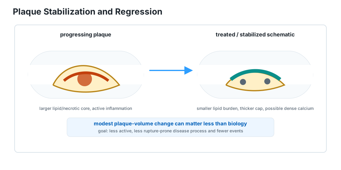
progressing plaque → slower or stopped progression → biological stabilization → sometimes modest reduction in plaque burden
Stabilization may matter more than shrinkage
A plaque's danger depends on more than how much space it occupies.
Important features include:
- lipid-rich/necrotic material
- inflammation
- fibrous-cap characteristics
- calcification
- tendency to rupture or erode
- resulting thrombosis risk
Intensive lipid-lowering treatment can change plaque composition as well as size.
Imaging studies of statin treatment have repeatedly found reductions in noncalcified/lipid-rich components and evidence of plaque stabilization.
Some calcified plaque can increase even while overall risk falls.
That sounds paradoxical until calcification is understood as one aspect of plaque remodeling rather than simply "more blockage."
LDL lowering and regression
Serial coronary-imaging trials have shown that intensive LDL lowering can produce modest average regression of coronary atheroma volume.
The effect is strongly related to the achieved LDL level and the magnitude of LDL reduction.
Additional LDL lowering with drugs such as ezetimibe or PCSK9 inhibitors can produce further favorable changes in selected high-risk populations.
The clinically important endpoint, however, is not winning a plaque-shrinkage contest.
It is reducing:
- myocardial infarction
- ischemic stroke
- cardiovascular death
- need for revascularization
Plaque imaging helps reveal mechanism; event reduction is what matters to patients.
Why established disease may not disappear
Advanced plaques contain more than cholesterol.
They can include:
- fibrous tissue
- calcium
- necrotic material
- remodeled vessel wall
Those structural changes may not fully reverse.
NHLBI therefore explicitly notes that severe atherosclerosis may not be fully reversible with currently available treatment.
Lifestyle matters even when plaque remains
Lifestyle interventions can improve several forces that drive atherosclerosis:
- blood pressure
- glucose control
- smoking exposure
- physical fitness
- body composition
- lipid profile
Medicines can target some of the same pathways more strongly or directly.
The useful conclusion is not that lifestyle alone can "wash plaque away."
It is:
changing the biological environment can make existing disease progress more slowly, become more stable, and sometimes regress measurably.
A useful distinction: lumen versus plaque
- The inside diameter of an artery is not a perfect proxy for total plaque burden.
- Early in atherosclerosis, the artery can remodel outward as plaque develops in the wall.
- Conversely, plaque composition can improve without a spectacular change in angiographic lumen diameter.
- This is another reason "percent blocked" is an incomplete picture of atherosclerotic biology.
Takeaway: Plaque is not immutable. Modern treatment can stabilize it and sometimes shrink it modestly, especially through intensive lowering of atherogenic lipoproteins. The realistic goal is a less active, less rupture-prone disease process and fewer cardiovascular events — not necessarily an artery returned to its teenage appearance.
Atherosclerosis is systemic
Plaques can occur in:
- coronary arteries
- carotid and cerebral circulation
- aorta
- renal arteries
- peripheral arteries in the legs
The clinical syndrome depends on where blood flow is impaired or where thrombosis/embolization occurs.
Risk factors act through multiple mechanisms
Major contributors include:
- elevated atherogenic lipoprotein exposure
- hypertension
- smoking
- diabetes
- kidney disease
- age
- inherited susceptibility
- inflammatory biology
These risk factors interact rather than acting as isolated switches.
Calcification is not the whole disease
Some plaques calcify as they evolve.
A coronary artery calcium scan can therefore be a useful marker of accumulated coronary atherosclerotic burden in selected risk-assessment contexts.
But calcium is a marker of plaque burden, not a complete map of every plaque's current danger.
Big idea: Atherosclerosis is an artery-wall disease. Chronic plaque formation creates the substrate; sudden clot formation often creates the emergency.