Stroke: Ischemic and Hemorrhagic
A stroke is acute brain injury caused either by:
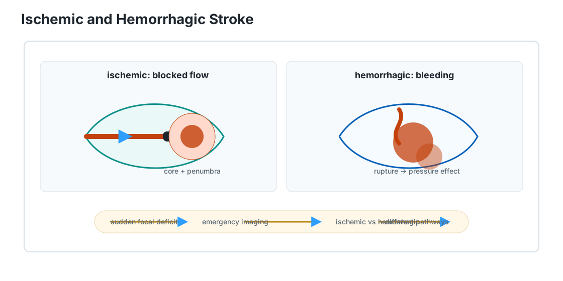
- loss of blood flow to part of the brain, or
- bleeding into or around the brain.
These are fundamentally different mechanisms, which is why brain imaging is urgently needed before treatment.
Ischemic stroke
An ischemic stroke occurs when an artery supplying brain tissue becomes blocked.
Major mechanisms include:
- local atherosclerotic thrombosis
- embolus from an artery such as the carotid
- embolus from the heart, especially in atrial fibrillation
- small-vessel occlusion
The result is:
arterial blockage
→ reduced cerebral blood flow
→ ATP failure
→ ion-pump failure
→ depolarization / excitotoxic injury
→ cell death if flow is not restored
The brain has high energy demand and little stored fuel, so prolonged interruption of flow is dangerous.
Core and penumbra
Severe ischemia can produce an irreversibly injured core.
Around it may be a penumbra: tissue with impaired function and reduced blood flow that is threatened but potentially salvageable.
This is why modern stroke treatment is organized around both time and imaging.
The old slogan "time is brain" is broadly correct, but modern treatment can sometimes identify salvageable tissue beyond simple clock cutoffs.
Acute treatment
For eligible ischemic-stroke patients, intravenous thrombolytic drugs such as alteplase or tenecteplase can dissolve clot.
Mechanical thrombectomy can physically remove selected large-vessel clots.
Treatment eligibility depends on timing, imaging, bleeding risk, anatomy and clinical circumstances.
It is not safe to infer treatment from symptoms alone because hemorrhage can mimic ischemic stroke.
Hemorrhagic stroke
A hemorrhagic stroke occurs when a blood vessel ruptures.
Bleeding can damage tissue directly and raise intracranial pressure.
Important forms include:
- intracerebral hemorrhage
- subarachnoid hemorrhage
Hypertension is an important risk factor for intracerebral hemorrhage.
Aneurysm rupture is an important cause of subarachnoid hemorrhage.
Why blood pressure is involved in both types
Chronic hypertension contributes to:
- atherosclerosis and small-vessel disease → ischemic stroke risk
- weakening/damage of small cerebral vessels → hemorrhagic stroke risk
So "stroke caused by high blood pressure" can refer to more than one mechanism.
Symptoms reflect anatomy
The brain is functionally organized.
Loss of blood flow in different territories can produce different deficits:
- face/arm/leg weakness
- sensory loss
- speech or language difficulty
- visual loss
- balance problems
- neglect or spatial deficits
Sudden focal neurological deficits are therefore a vascular warning pattern.
The American Stroke Association's current recognition mnemonic is BE FAST:
- Balance
- Eyes
- Face
- Arm
- Speech
- Time
The important action is not to diagnose the stroke subtype at home. It is to get emergency assessment quickly.
TIA
A transient ischemic attack (TIA) produces transient focal neurological symptoms from ischemia without the same completed infarction pattern.
Symptoms resolving does not make the event trivial.
A TIA can be a warning of high near-term stroke risk and requires urgent medical evaluation.
Big idea: "Stroke" describes an acute brain injury syndrome, not one single plumbing problem. Ischemic stroke is blocked flow; hemorrhagic stroke is bleeding; imaging distinguishes them because treatment can be opposite.